Jim's Study (where it all happened)
One or two visitors to this
web-site have asked where/how the images are made, so I've created a virtual
tour of my study. Be patient while the images download.
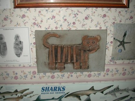
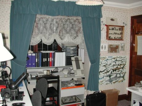
First of all my favourite fossil of a very rare cat
from the old Czech republic (SEM image of fossilised weevle from Sheppey
to it's right (think about it) and a Sheppey starfish to it's left)
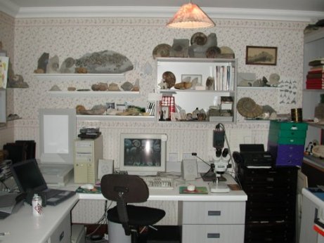
panning round the room to the right, bookcases &
display cabinet and an old Russian microscope with amazing optics.
moving across, Kaiser copy stand with "shadow-box"
and Schott fibre-optic light source behind. You have to imagine a Nikon
990 CoolPix camera mounted to the stand (I'm taking these pictures with
it). This is where my macro-photography takes place. Take a look at the
bivalves section on the Gault
web-site for examples of the imagry.
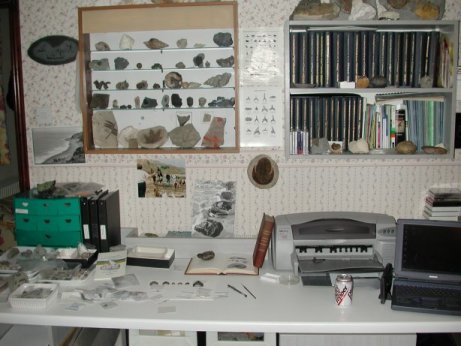
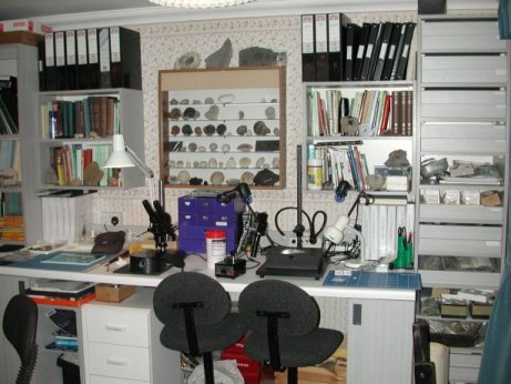
moving right, another working area. Folders with literature,
scans etc
right again, another working area. Copy of new book
on Sheppey
Fossils in background. Then colour HP printer with a set of the "Treatise
on Invertebrate Paleontology" in the bookcase behind; and back to
the Dr. Pepper completing the circuit.
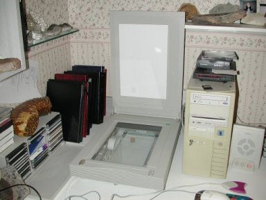
Close-up of HP Scanner - where most of the ammonite
images have been made. Mammoth tooth on top of CD box. Images also made
with the Nikon.
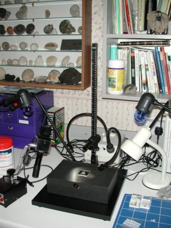
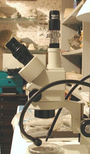
Meiji EMZ-TR Trinocular microscope with relay lens
and adapters for the Nikon CoolPix 990 Digital Camera used for creating
images on the pages on the Gault Foraminiferida
and Ostracoda and elsewhere
on this web-site. If you would like to know more about the connections
between the microscope and camera go to adapters.
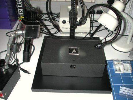
This is my "shadow-box" placed
on a Kaiser Copy Stand and also used with the Nikon 990. The box is lined
with matt black card, a square window cut out and glass inserted with
a millimeter measuring scale on the bottom edge. Schott fibre-optics unit
in background. Fossil placed on window and image made in macro-mode.
Acknowledgement: Special thanks
is due to David Ward of Orpington for introducing me to the Nikon camera
and for help with ideas and techniques for making digital images, including
use with the Meiji microscope and copy-stand.